Minimally Invasive Spine Surgeon

Minimally Invasive Spine Surgeon In Nashik

Dr. Shekhar Chirmade is Minimally Invasive Spine Surgeon In Nashik. Spine surgery is typically recommended only when a period of nonsurgical treatment. such as medications and physical therapy. has not relieved the painful symptoms, caused by the back problem.

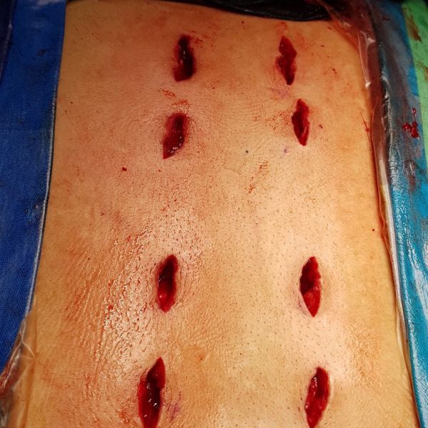
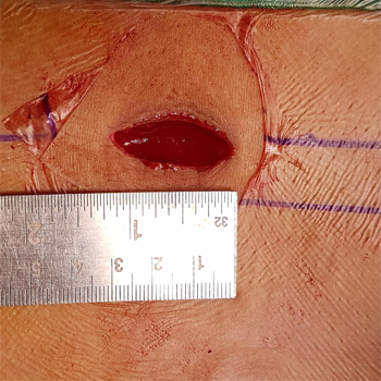
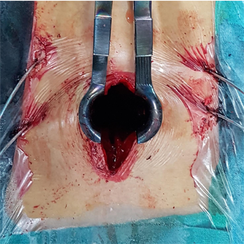
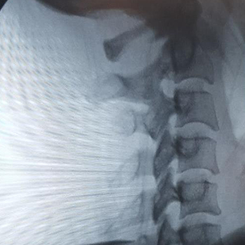
Surgery is only considered if the spine surgeon can pinpoint the exact source of pain, such as a herniated disk or spinal stenosis. Spine Surgery is traditionally done as “open surgery,” meaning the area being operated on is opened with a long incision to allow the surgeon to view and access the anatomy.

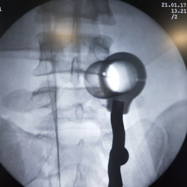
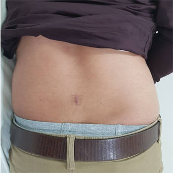
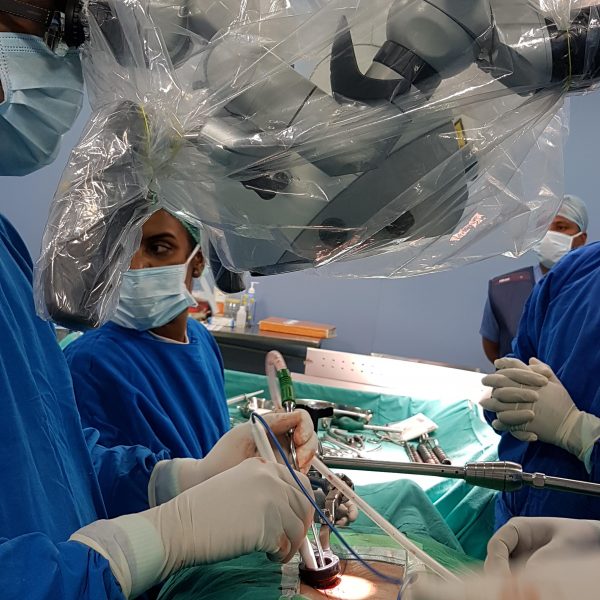
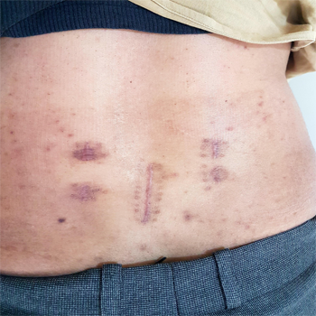
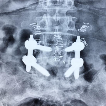
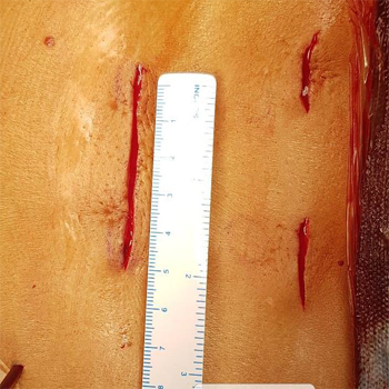
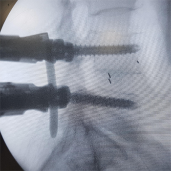
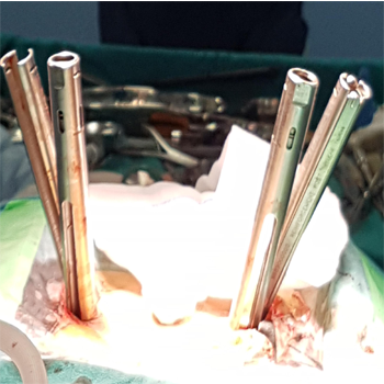
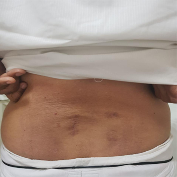
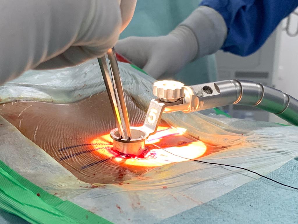
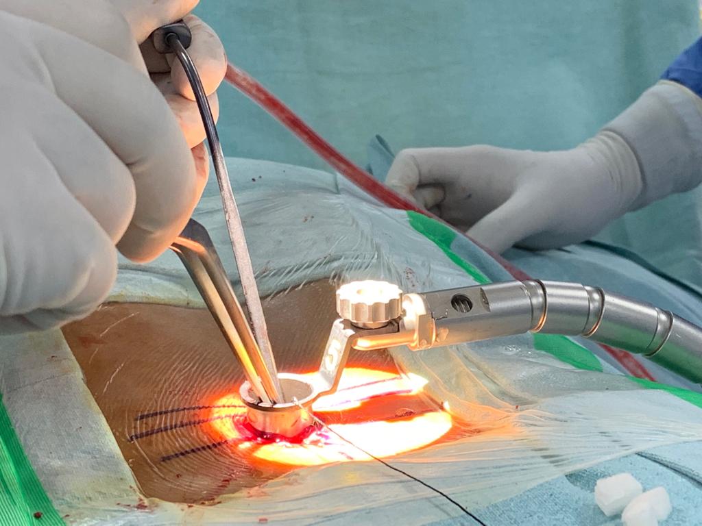
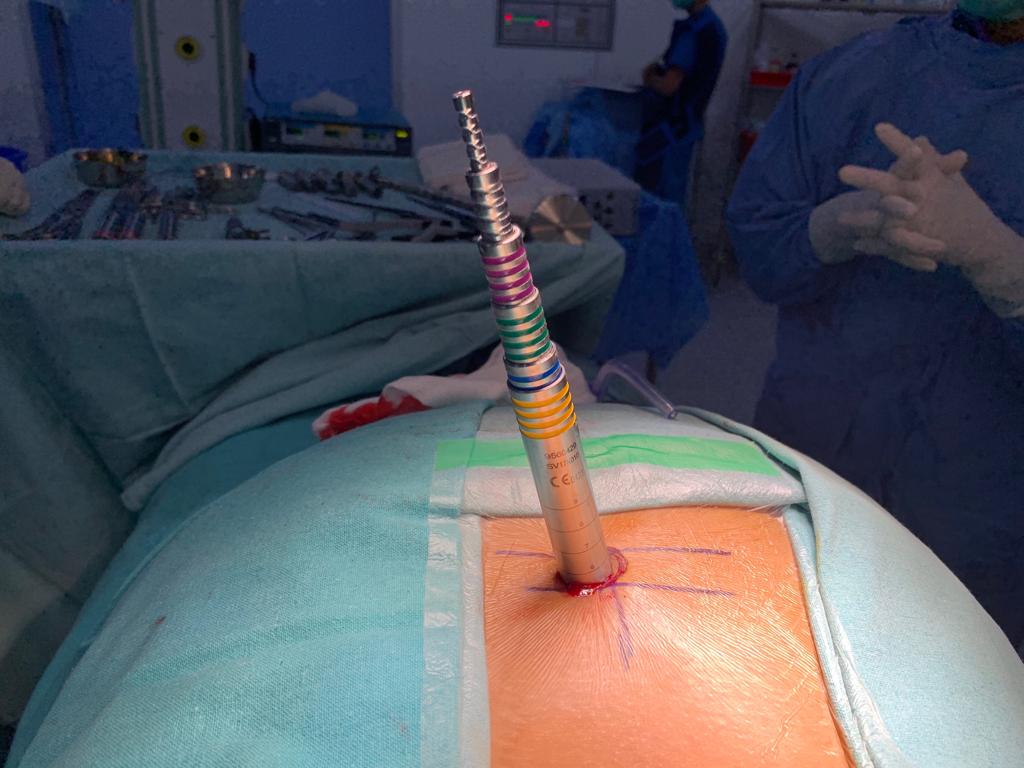
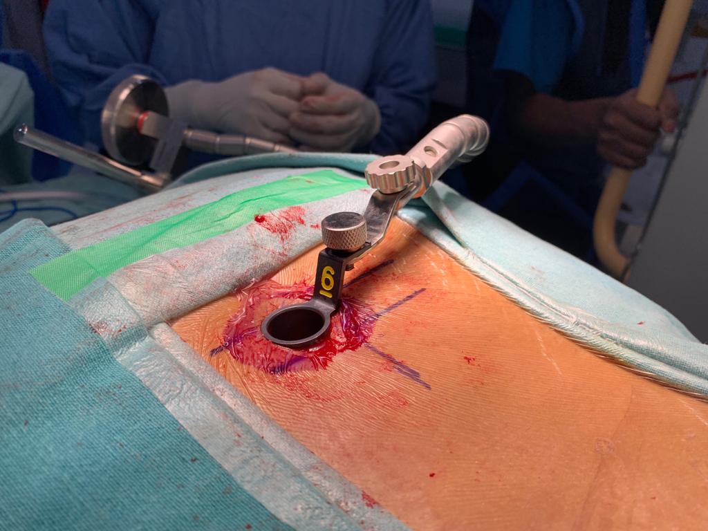
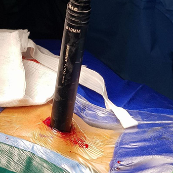
However, technological advances have allowed more back and neck conditions. to be treated with a Minimally Access Surgical Technique (MAST) or Minimally Invasive Spinal Surgery (MISS). Minimally Access Techniques are beginning to be used for a wider range of spine procedures, and have been used for common procedures. like decompression and spinal fusion.

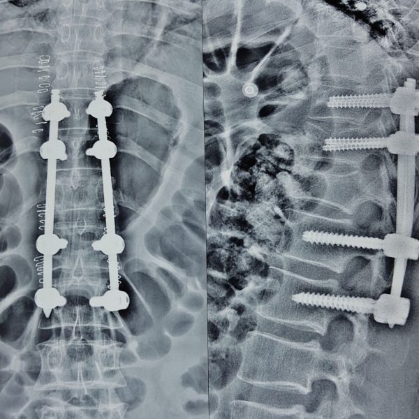
corrects problems with the small bones of the spine (vertebrae). The basic idea is to fuse together the painful vertebrae so that they heal into a single. solid bone As opposed to open spine surgery, Minimally Invasive Surgical approaches can be faster, safer and require less recovery time.

Because of the reduced trauma to the muscles and soft tissues, the potential benefits are:

  • Better cosmetic results from smaller skin incisions. (sometimes as small as 2 centimeters).
  • Less blood loss from surgery.
  • Reduced risk of muscle damage since less or no cutting of the muscle is required.
  • Reduced risk of infection and postoperative pain.
  • Faster recovery from surgery and less rehabilitation required.
  • The decreased requirement of pain medications after surgery.

Performing the surgery using a tubular retractor:

This technique involves progressive dilation of the soft tissues, as opposed to cutting directly through the muscles. By using tubes to keep the muscles out of the way. the surgery can be performed through the incision without having to expose the area widely with the help of endoscopic or microscope.

focused down the tube to assist with performing the surgery through a minimal access strategy. Once the procedure is complete, the tubular retractor can be removed. allowing the dilated tissues to come back together. Depending on the extent and type of surgery necessary, incisions can often be small.

Conditions Treated Using MIS Procedures.

    • Herniated disc.
    • Lumbar spinal stenosis.
    • Spinal infections.
    • Spinal instability.
    • Vertebral compression fractures.
    • Spinal Tumors.

A number of methods can be used to minimize trauma during MIS surgery. Some of the more common techniques include:

Percutaneous placement of screws and rods:

Depending on the condition of the patient, it may be necessary to place instrumentation. such as rods and screws, to stabilize the spine or to immobilize the spine to facilitate fusion of the spinal bones. Traditional approaches for placement of screws require extensive removal of muscle and other tissues from the surface of the spine.

    • However percutaneous means through the skin typically involves inserting rods and screw through relatively small skin incisions without cutting or dissecting the underlying muscle.
    • With the aid of X-ray images, guidewires are placed through the skin and into the spinal vertebrae along the desired paths for the screws.
    • Then, screws are placed over the guide wires and follow the path of the wires. These screws have temporary extenders that extend outside of the skin and subsequently removed after helping to guide passage of rods to connect and secure the screws.

MISS-Microlumbar Discectomy:

Spinal discs are essentially elastic rings with soft material inside that serve as cushions between the vertebral bones. If the elastic ring becomes incompetent or weakened. the soft tissue inside can extrude or herniate outside of the elastic ring.

The herniated disc material can compress the nerves passing by, thus causing pain. If surgical treatment is recommended to trim or remove the herniated disc. it may be possible to perform this procedure with MIS surgery using tubular dilators and a microscope or endoscope.

Spinal decompression:

Spinal stenosis, which is a narrowing of the vertebral column, is a common condition. that can result in compression of the nerves. This can produce a variety of symptoms, including pain, numbness and muscle weakness.

If surgery is recommended, it may be possible to remove the bone and soft tissues causing the nerve compression. through an MIS approach using tubular dilators and a microscope or endoscope. The more common decompressive procedures include laminectomy and foraminotomy.

MIS Fusion Procedure

  • Minimally Invasive Posterior Lumbar Interbody Fusion (PLIF)
  • Minimally Invasive Transforaminal Lumbar Interbody Fusion (TLIF)

This is an MIS technique that is performed in patients with refractory mechanical low back and radicular pain associated with spondylolisthesis, degenerative disc disease, and recurrent disc herniation.

The TLIF approach may also have potential in patients with low back pain caused by postlaminectomy instability, spinal trauma or for treating pseudoarthrosis. The procedure is performed from the back (posterior) with the patient on his or her stomach.

Posterior Cervical Foraminotomy.

The typical patient presents with pain down one arm which may radiate to the hand. Cervical disc problems are exceedingly common and it is important to realize that in the vast majority of cases non-operative management works very well. Most patients settle within 6-12 weeks after the onset of symptoms.

Posterior cervical foraminotomy is a minimally invasive procedure designed to enlarge to space. through which the nerve root exits from the spinal cord. (the so-called neural foramen) and at the same time try to remove any piece of a disc.

which is pushing on the nerve. Usually, a small window is made on one side of a spinous process, at the junction of the lamina and facet joint. through the removal of some bone and ligament to allow visualization of the involved root.

Using a high-speed drill and microinstruments, once the nerve root affected is identified. the whole out of which the nerve passes is enlarged.

Scroll to Top